. 2015 Jul 8;86(2):206–213. doi: 10.2319/022515-118.1

Figure 2.

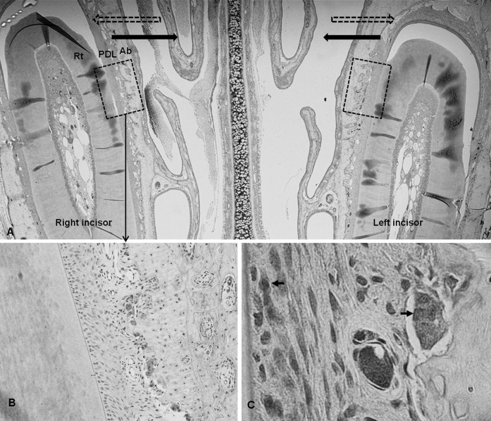
Figure 2. Microphotograph of immunohistochemical staining in the experimental groups. (A) Representative microphotograph of the whole observation area, including teeth and periodontal tissues used in the experiment, ×20. Dashed arrow indicates direction of previous orthodontic tooth movement; solid arrow, direction of relapse movement. Former tension sides were changed into compression sides. Dashed box indicates the range of interest. (B) Immunoreactivity to MMPs was seen in the multinucleated cells along the resorbed bone surface, ×200.(C) Immunoreactive cells, ×1000. Rt indicates incisor root; PDL, periodontal ligament; Ab, alveolar bone.

Microphotograph of immunohistochemical staining in the experimental groups. (A) Representative microphotograph of the whole observation area, including teeth and periodontal tissues used in the experiment, ×20. Dashed arrow indicates direction of previous orthodontic tooth movement; solid arrow, direction of relapse movement. Former tension sides were changed into compression sides. Dashed box indicates the range of interest. (B) Immunoreactivity to MMPs was seen in the multinucleated cells along the resorbed bone surface, ×200.(C) Immunoreactive cells, ×1000. Rt indicates incisor root; PDL, periodontal ligament; Ab, alveolar bone.