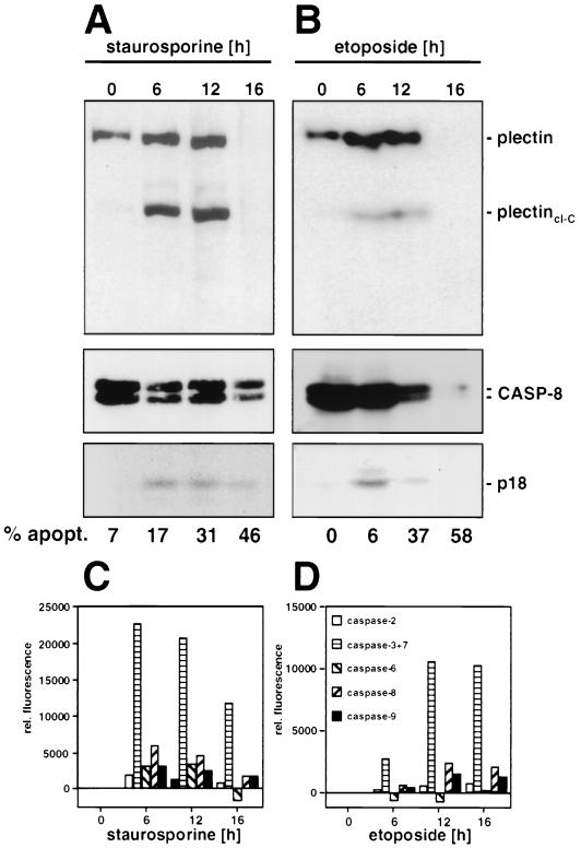
FIG. 5.
Cleavage of plectin during drug-induced apoptosis. Jurkat cells were incubated with staurosporine (1 μM) (A) or etoposide (20 μg/ml) (B) for the indicated time periods. Analysis of plectin and caspase 8 cleavage fragments was done as described for Fig. 4A and B; quantification of the apoptotic cells was done as described in Materials and Methods. (C and D) Caspase activities during drug-induced apoptosis, determined as described in the legend for Fig. 4C and D.

