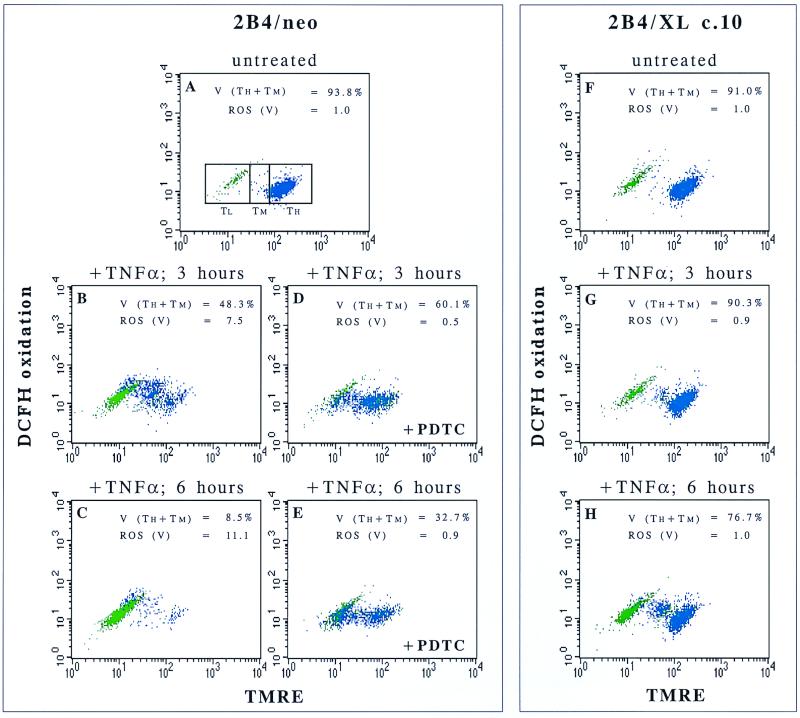
FIG. 5.
Following TNF-α treatment, ROS are induced after a partial decrease in ΔΨm and are important mediators of mitochondrial membrane depolarization. Control cells (2B4/neo) and Bcl-xL-expressing cells (2B4/Xl c.10) were either left untreated (A and F) or treated with TNF-α (B to E, G, and H). PDTC, a ROS scavenger, was added to 2B4/neo cells (D and E). At the indicated time points, cells were doubly stained with TMRE and DCFH-DA to analyze the increase in ROS levels in parallel with loss of ΔΨm. Cells were divided by forward-scatter and side-scatter criteria into live cells (blue) and dead cells (green). Other subgroups were defined by ΔΨm levels: TH, TMRE high; TM, TMRE medium; TL, TMRE low. Viability (V) was defined in this assay as the percentage of cells in the TH + TM subgroups. ROS levels (fold above untreated cells [see Materials and Methods]) were analyzed for viable cells only [ROS (V)].

