. 2015 Apr 3;86(1):66–73. doi: 10.2319/121414.1

Figure 1.

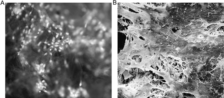
Figure 1. Microscopic observations of PDLtm. (A) Scaffolded, spindle-shaped PDLCs growing densely, with green-yellow nuclei and orange-red cytoplasm, under fluorescence inverted microscope after arcidine orange staining (×200). (B) Integration of PDLCs, secreted extracellular matrix, and PLGA scaffolds observed under SEM (×1200).

Microscopic observations of PDLtm. (A) Scaffolded, spindle-shaped PDLCs growing densely, with green-yellow nuclei and orange-red cytoplasm, under fluorescence inverted microscope after arcidine orange staining (×200). (B) Integration of PDLCs, secreted extracellular matrix, and PLGA scaffolds observed under SEM (×1200).