. 2015 Mar 26;86(1):74–80. doi: 10.2319/111914-830.1

Figure 1.

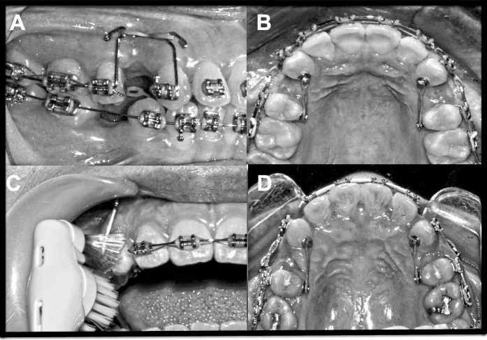
Figure 1. (A, B) Illustration of the mechanics employed for canine distalization; 60 g force was applied to both the control and experimental canines. (C) Application of vibratory stimuli to the canine on the experimental side using an electric toothbrush. (D) Images of the experimental (left) and control (right) sides in a representative case at T3.

(A, B) Illustration of the mechanics employed for canine distalization; 60 g force was applied to both the control and experimental canines. (C) Application of vibratory stimuli to the canine on the experimental side using an electric toothbrush. (D) Images of the experimental (left) and control (right) sides in a representative case at T3.