
Figure 3. Early postnatal differentiation of Sertoli and germ cells occurs normally in cKO testes.
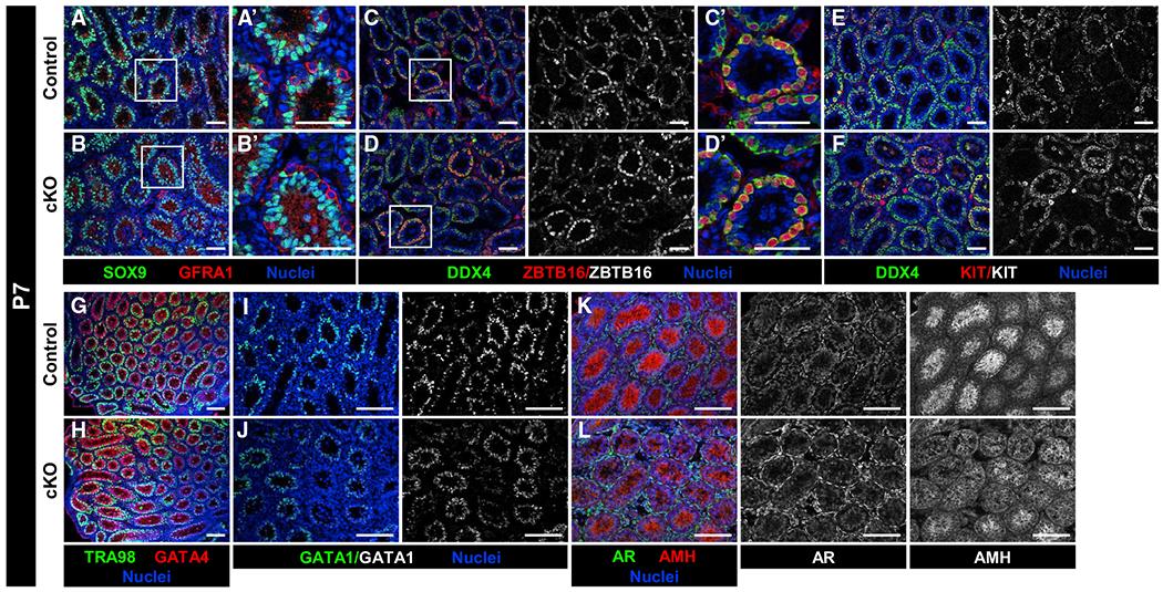
Immunofluorescence images of P7 control Dhh-Cre;Cdc42flox/+ (A, C, E, G, I, and K) and cKO (B, D, F, H, J, and L) testes. (A’)–(D’) are higher-magnification images of the boxed regions in (A)–(D).
(A–H) Compared to P7 controls (A, C, E, and G), P7 cKO testes (B, D, F, and H) have similar numbers and localization of Sertoli cells (SOX9+, GATA4+), undifferentiated spermatogonia (GFRA1+, ZBTB16+), differentiating spermatogonia (KIT+), and overall germ cells (DDX4+, TRA98+).
(I and J) GATA1 expression is detected in both control (I) and cKO (J) Sertoli cells.
(K and L) Both AMH and AR are detected within control (K) and cKO (L) testes, but AMH expression appears slightly decreased in a subset of cKO Sertoli cells.
Scale bars, 100 μm.