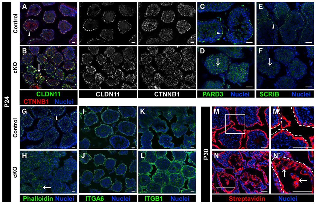
Figure 6. Loss of Cdc42 leads to disruptions in Sertoli cell polarity and BTB function in cKO juvenile testes.

Immunofluorescent images of P24 control Dhh-Cre;Cdc42flox/+ (A, C, E, G, I, and K), P30 control (M), P24 cKO (B, D, F, H, J, and L), and P30 cKO (N) testes. (M’) and (N’) are higher-magnification images of the boxed regions in (M) and (N).
(A–H) While control (A, C, E, and G) testes showed compartment-specific enrichment of CLDN11, CTNNB1, PARD3, SCRIB, and phalloidin staining (arrowheads), cKO testes (B, D, F, and H) exhibited aberrant localization of these factors (arrows).
(I–L) Both control (I and K) and cKO (J and L) testes displayed strong expression of ITGA6 and ITGB1 in the tubule basement membrane; however, cKO testes regularly displayed ectopic expression of ITGB1 in the central portion of tubules.
(M and N) While P30 control (M) testes showed only the interstitial and basal tubular presence of biotin (detected by streptavidin staining) in a tracer assay, cKO testes (N) showed widespread presence of biotin deep within tubules (arrows in N’) (n = 3 testes each for controls and cKO). Dashed lines indicate tubule boundaries.
Scale bars, 50 μm.
