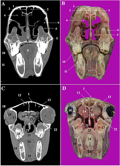
FIGURE 5.

CT scan (a, c) and cross‐sectional (b, d) images of the middle and caudal nasal cavity at the level of the orbit in the Arabian horse. 1: Nasal bone, 2: frontal septum between right and left frontal sinus, 3: rostral frontal sinus, 4 conchofrontal sinus, 5: rostral maxillary sinus, its lateral compartment, 6: medial compartment of rostral maxillary sinus, 7: nasal septum, 8: hard palate, 9: tongue, 10: 1st upper molar tooth (Triadan 109 and 209), 11: 1st lower molar tooth (Triadan 309 and 409), 12: medial frontal sinus, 13: caudal frontal sinus, 14: ethmoidal labyrinth, 15: middle nasal concha, 16: eye, 17: periorbital fat and ocular muscles, 18: lens, 19: masseter muscle, 20: 2nd lower molar tooth (Triadan 310 and 410), 21: nasopharynx, 22: caudal maxillary sinus, 23: soft palate, 24: root of the tongue, 25: omohyoid muscle, 26: 3rd upper molar tooth (Triadan 111 and 211)
