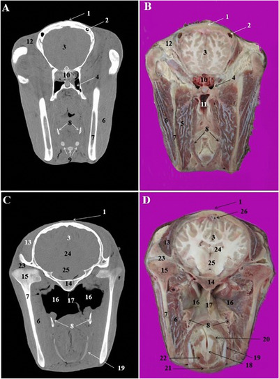
FIGURE 6.

CT scan (a, c) and cross‐sectional (b, d) images of the middle nasal cavity at the level of the brain in the Arabian horse. 1: Frontal bone, 2: most caudal part of the frontal sinus, 3: frontal lobe of brain, 4: sphenopalatine sinus, 5: medial pterygoid muscle, 6: masseter muscle, 7: ramus of the mandible, 8: stylohyoid bones, 9: laryngeal muscles, 10: ethmoidal labyrinth, 11: pharyngeal recess, 12: retro‐orbital fat, 13: temporalis muscle, 14: body of basisphenoid bone, 15: condylar process of the mandible, 16: guttural pouch, 17: septum of guttural pouch, 18: arytenoid cartilage, 19: thyroid cartilage, 20: arythenoideous transversous, 21: cricothyroid muscle, 22: thyroarythrnoid muscle, 23: temporomandibular joint, 24: thalamus, 25: hypothalamus, 26: dorsal sagittal sinus of dura mater
