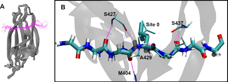
Fig 2. Conserved βSBD-substrate binding conformation.
(A) Overlay of 18 bound substrate structures (S1 Table), aligned using the backbone atoms of the βSBD. The SBDs are show in gray cartoons, and the substrates in magenta backbone traces. Only the forward orientation substrates are included. (B) Overlay of two forward and reverse orientation peptide substrates (forward: NRLLLTG, PDB: 1DKZ [10]; reverse: NRLILTG, PDB: 4EZY [19]; underlines indicate residues at Site 0). The forward orientation is shown in thick bonds and the reverse in thin bonds. The backbone hydrogen bonds between βSBD and the substrate are shown in magenta dashed lines. Note that backbone hydrogen bonds involving M404, S427 and A429 completely overlap in two orientations. Side chains are shown only for the central L at Site 0. For more viewing angles of panel B, see S1 Movie.

