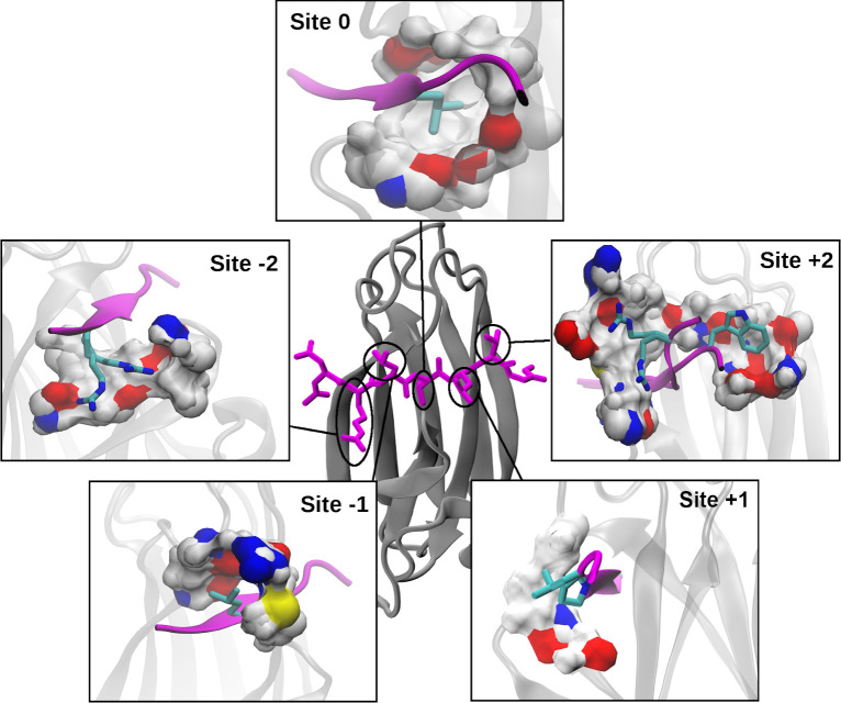
Fig 3. Substrate-interacting sites of DnaK.
The center image shows a NRLLLTG substrate (magenta sticks) bound to βSBD (gray cartoon) (PDB: 1DKX). Each zoom-in shows the surface of one of the five binding sites on DnaK, colored by atom type: O (red), N (blue), C/H (white), S (yellow). The side-chain conformations shown were selected to clearly illustrate the extent of each site. These additional side-chain and substrate backbone conformations are taken from: (Site -2) 4JWD, (Site -1) 1DKX, (Site 0) 1DKX (for more viewing angles, see S2 Movie), (Site +1) 1DKZ, 4F00 and (Site +2) 4JWI, 4EZN, 4EZO. The binding site surface was generated using DnaK residues within 5 Å of the substrate side chain(s) at each site, with some atoms excluded for clarity.

