Figure 1.

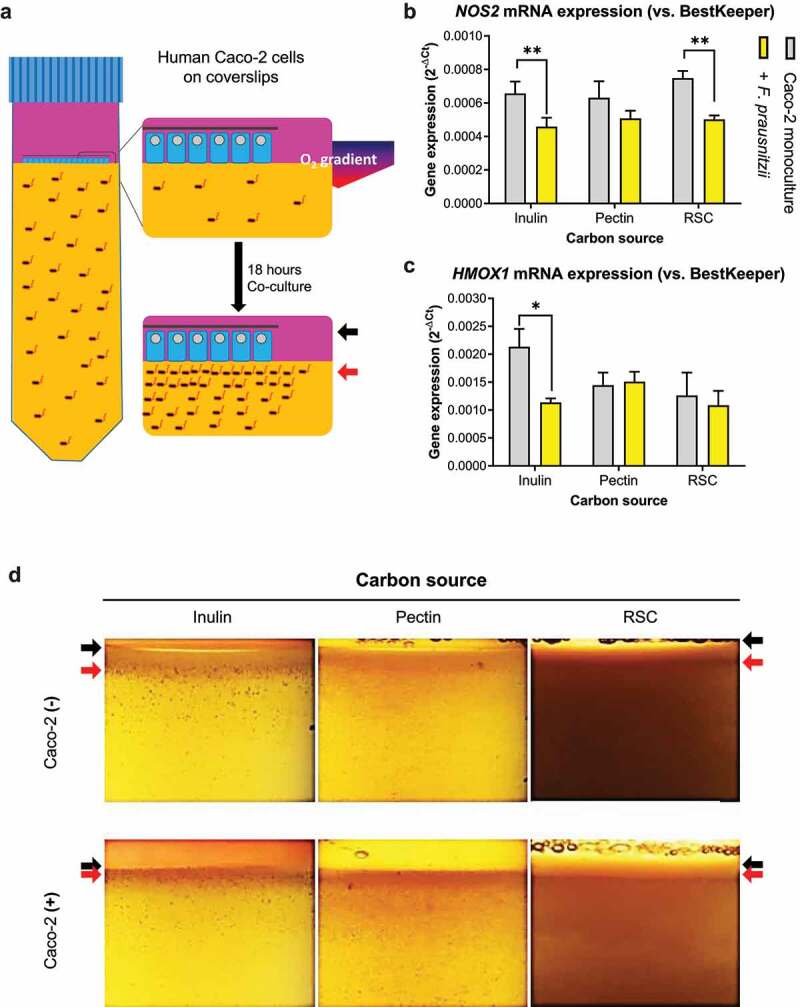
Metabolism of prebiotics by F. prausnitzii decreases inflammatory markers in Caco-2 cells. (a) Schematic representation of the HoxBan system, showing the oxic-anoxic interphase created between the Caco-2 monolayer (in human compartment, pink colored) and bacterial compartment (yellow color, representing the YCFA medium). The zoom schematically shows rim formation after 18-hour co-culture, where black arrows indicate the coverslip and red arrows indicate bacterial rim localization. (b-c) Effect of different prebiotic carbon sources on F. prausnitzii-regulated expression of NOS2 (inflammation marker, B) and HMOX1 (oxidative stress marker, C) in Caco-2 cells. Especially inulin-grown F. prausnitzii suppresses NOS2 and HMOX1 expression. (d) Prebiotic-grown F. prausnitzii in the absence and presence of Caco-2 cells. In the absence of Caco-2 cells (top panels), F. prausnitzii growth is enhanced (red arrow) in the upper part of the bacterial compartment forming a rim below the oxic-anoxic interphase (black arrow). In the presence of Caco-2 cells (bottom panels), F. prausnitzii growth is further enhanced (red arrow) and colonies appear closer to the oxic-anoxic interphase where the Caco-2 cells reside (black arrow). All experiments were performed with two biological replicates, each with an N = 3. *P < .05; **P < .01
