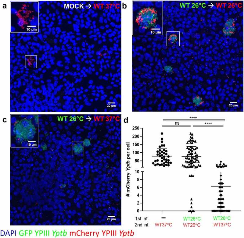
Figure 7.

M cells infected with WT 26°C have reduced ability to be reinfected by WT 37°C. (a-d) Differentiated HIE25 RT+ ileal monolayers were (a) mock infected with noninfectious media, washed, and infected for 2.5 hours with 5 × 10⁶ CFU of WT 37°C expressing mCherry (red), or (b) infected for 2.5 hours with 5 × 10⁶ CFU of WT 26°C expressing GFP (green), washed, and infected for 2.5 hours with 5 × 10⁶ CFU of WT 26°C expressing mCherry (red), or (c) infected for 2.5 hours with 5 × 10⁶ CFU of WT 26°C expressing GFP (green), washed, and infected for 2.5 hours with 5 × 10⁶ CFU of WT 37°C expressing mCherry (red). Monolayers were stained with DAPI (nuclei-blue). XY planes are maximum intensity projections. Magnified insets of an M cell are shown in upper left corner. (d) The number of mCherry Yptb per cell were counted using Volocity software. Each point represents an infected M cell. Bars indicate mean and SD. Data were pooled from 3 independent experiments with 3 fields analyzed per Transwell and averaged. Statistics were performed using a one-way ANOVA and Tukey’s post hoc multiple comparison tests
