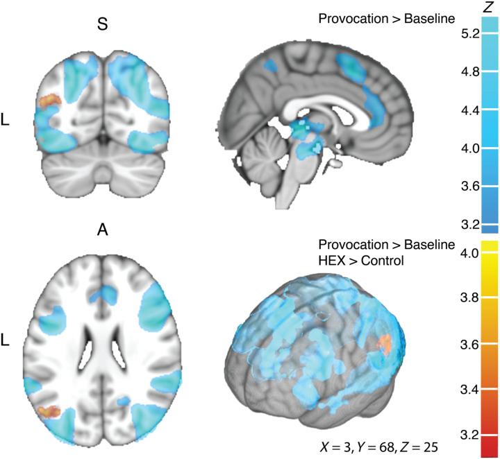
Fig. 6. Provocation recruited an extensive brain network.
The group image for both women and men (n = 49), in coronal (top left), sagittal (top right), axial (bottom left), and surface (bottom right) views. In all panels, shades of blue reflect the Provocation > Baseline contrast, and shades of orange reflect the Provocation > Baseline with an additional HEX > Control contrast. In blue, provocation induced activity in the fusiform gyrus, OFC, insula, superior temporal gyrus, anterior cingulate cortex, inferior frontal gyrus, pre-SMA, precuneus, ventral tegmental area, periaqueductal gray area, and thalamus (for full list, coordinates, and peak activation, see table S2). In orange, HEX induced activation in left angular gyrus (AG). Z statistic images were thresholded using clusters determined by Z > 3.1 and a (corrected) cluster significance threshold of P = 0.05 (as described in Materials and Methods).

