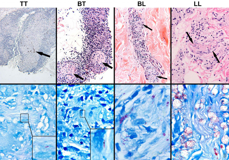
Fig. 1.
Inflammation and infection of cutaneous nerves across the leprosy spectrum. The inflammatory responses in and around cutaneous nerves are shown in the upper panel; arrows highlight recognizable nerve twigs. The immunopathologic classifications of leprosy, TT to LL, are indicated at the top of the figure (see text; mid-borderline, BB, is not shown). The TT lesion (upper left) is composed of a well-organized epithelioid granuloma that has nearly destroyed the nerve, remnants of which are shown by S-100 staining. The granulomatous inflammatory response becomes less organized across the spectrum until, at the LL extreme, it is composed of disorganized aggregates of foamy histiocytes, seen here surrounding a nerve (upper right). (TT, S-100, original magnification × 10; BT, BL, LL, hematoxylin–eosin, original magnification × 250). The demonstration of acid-fast bacilli within nerves is pathognomonic of leprosy. In the lower panel, Fite-stained sections reveal the corresponding intensity of M leprae infection in cutaneous nerves across the spectrum. M. leprae are rare and difficult to demonstrate in nerves of TT and BT lesions; they have been photographically enlarged in the insets. In contrast, bacilli are abundant and easily recognized in BL and LL lesions. (Fite/methylene blue, original magnification × 1000.) (From Scollard et al., The Continuing Challenges of Leprosy. 2006. Clinical Microbiology Reviews, 19: 338–381

