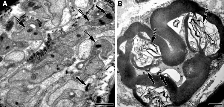
Fig. 2.
Electron micrographs of Mycobacterium leprae–infected peripheral nerves of Armadillos. A Electron micrograph of a M. leprae-infected tibial nerve section containing Remak bundles (unmyelinated axons) with M. leprae (arrows) in axoplasm and Schwann cell cytoplasm. Many Schwann cell process are denervated (broken arrows) but contain M. leprae. Scale bar: A = 1 µm. B Cross section of a myelinated axon with M. leprae within axoplasm (arrows). The nerve shows extensive demyelination (broken arrows) and axonal disruption. Scale bar: B = 2 µm

