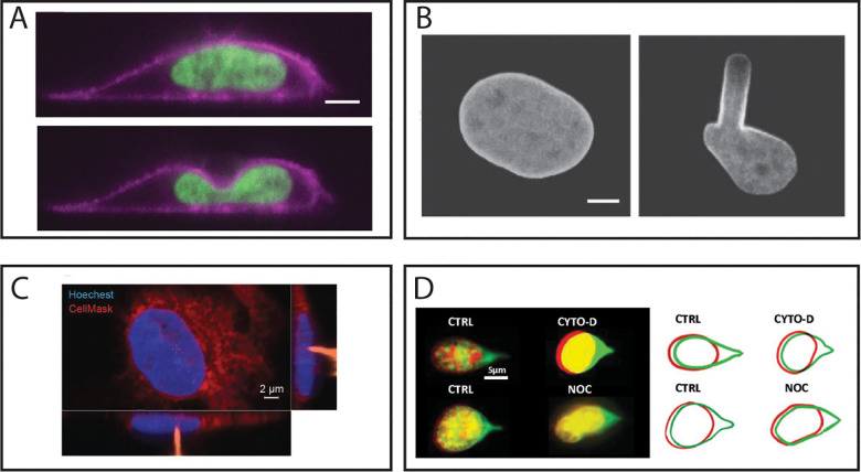
FIG. 3.
Considerations in the geometry of nuclear perturbation. (a) Side-view images of a nucleus being compressed by an AFM with a 6 μm diameter beaded tip, as visualized in Hobson et al. (b) Nuclear deformation during an MA experiment, as visualized in Rowat et al. (c) Wang et al. used AFM to show inhibition of the actin cytoskeleton reduced the viscoelastic response of the nucleus. Images show the nucleus (blue) being perturbed via a sharp AFM tip (orange) from above. (d) Neelam et al. used single-pipette micromanipulation to show inhibition of the actin cytoskeleton did not alter the nuclear response to external force. Red and green images and outlines represent nuclear shape before and after stretching with a micropipette. Images from (a) are reproduced with permission from Hobson et al., Mol. Biol. Cell 31(16), 1788–1801 (2020). Copyright 2020 Authors, licensed under a Creative Commons Attribution (CC BY) license. Images from (b) are reproduced with permission from Rowat et al., Biophys J. 91(12), 4649–4664 (2006). Copyright 2006 Elsevier. Images from (c) are republished with permission from Wang et al., J. Cell Sci. 131(13), jcs209627 (2018). Copyright 2018 The Company of Biologists Ltd., Clearance Center, Inc. Images from (d) are reproduced with permission from Neelam et al., Proc. Natl. Acad. Sci. U. S. A. 112(18), 5720–5725 (2015). Copyright 2015 National Academy of Sciences.

