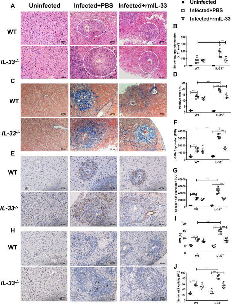
Figure 2.
IL-33 deficiency aggravated liver pathological lesions in murine schistosomiasis japonica. WT and IL-33−/− mice were divided into uninfected group, infected plus rmIL-33 group and infected plus PBS group. Each mouse in the infected groups was infected with 20 cercariae through shaved abdominal skin. Mice in the infected plus rmIL-33 group were intraperitoneally injected with exogenous rmIL-33 (dissolved in sterile PBS solution) from the 4th week to 8th week post infection, with the total 5 μg of rmIL-33 per mouse. The mice in the infection plus PBS group were simultaneously given the equal volume of PBS. The liver weight and body weight of all mice were recorded. At the 8th week post infection, all mice were sacrificed and the liver and peripheral blood were collected. (A) Representative images of HE staining of liver sections (single egg granuloma indicated by white circles) and (B) the statistical graphs of single granuloma area. (C) Representative images of Masson’s staining of liver sections and (D) the statistical graphs of the blue collagen area proportion. The hepatic expression of two main extracellular matrix components, α-SMA (E) and Collagen 1a1 (H). The statistics of IOD of α-SMA (F) and Collagen 1a1 (G). The statistical graphs of HMI (Hepatic Mass Index, (I)) and ALT (Alanine aminotransferase, (J)) levels in peripheral blood from mice. Data are expressed as means ± SEMs based on 6 mice in each group and from 2 independent experiments. Asterisks mark significant differences among different groups (**P < 0.01, ***P < 0.001).

