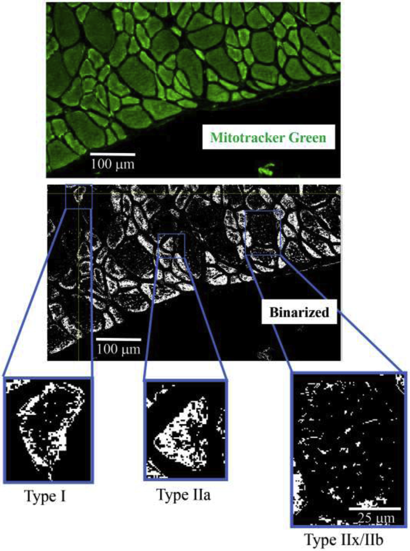
Figure 2:

Mitochondria in DIAm fibers were labeled using MitoTracker Green (upper photomicrograph) in alternate serial sections (same fibers used for fiber type classification and SDHmax measurements) cut at 10 μm thickness. MitoTracker Green fluorescence was visualized using Olympus FV2000 laser scanning confocal microscope and fluorescence intensity thresholded to create binary images of labeled mitochondria (middle photomicrograph). Representative binarized images of mitochondria in type I, IIa and IIx/IIb DIAm fibers are shown in the lower photomicrographs.
