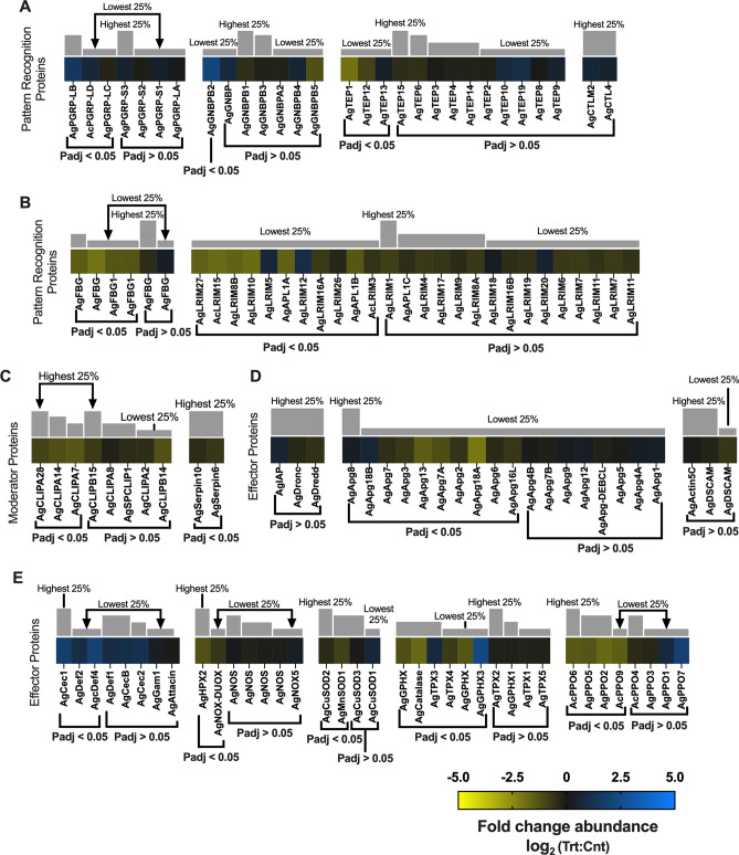
Figure 10.
Immune response differential transcript abundances following relevant sporozoite infections. Immune response transcripts that were present at significantly higher (blue) or lower (yellow) levels in Plasmodium falciparum sporozoite infected treatments; non-differentially expressed immune response transcripts are denoted as zeros (black). Immune response genes within each group organized by adjusted p-value and subsequently arrayed left to right from most abundant to least abundant based on FPKM values (quartile bars above each image). (A, B) Pattern recognition proteins. (C) Modulator proteins. (D, E) Effector proteins. Log2 scale indicates transcript abundances that were significantly higher (blue) or lower (yellow) in sporozoite infected treatments (Trt) vs. naïve controls (Cnt).

