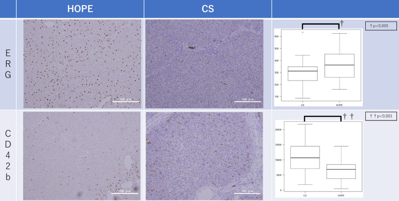
Figure 6.
Immunohistochemistry. At the end of reperfusion, the number of anti-ERG staining-positive SEC nuclei counted automatically by ImageJ in the HOPE group (360.54 ± 118.87 per field) was significantly higher than that in the CS group (285.13 ± 107.87 per field) (P = 0.002). At the end of reperfusion, the positive area of anti-CD42b staining counted automatically by ImageJ in the HOPE group (6317.06 ± 3235.14 per field) was significantly smaller than that in the CS group (10,761.50 ± 5643.620 per field) (P < 0.001). CS cold storage, HOPE hypothermic oxygenated machine perfusion preservation, SEC sinusoid epithelial cells.

