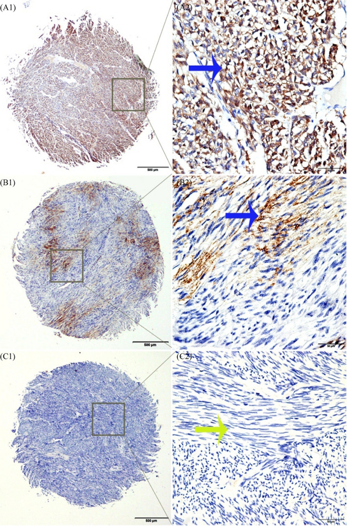
FIGURE 1.

Immunohistochemical staining of FABP4 in clinical tissue samples of GISTs. A1: High cytoplasmic staining of FABP4 in the tissue microarray samples. A2: Specific high positive staining for FABP4 in the cytoplasm. B1: Low cytoplasmic staining of FABP4 in gastrointestinal stromal tumor tissues. B2: Specific low positive staining for FABP4 in the cytoplasm. C1 and C2: Negative staining for FABP4. Original magnification: A1, B1, C1 × 40; A2, B2, C2 × 400
