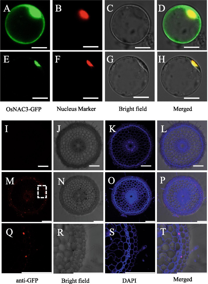
Fig. 2.
Subcellular and cellular localization of OsNAC3. (A-H) Rice protoplast co-expressing GFP-OsNAC3 or GFP with OsGhd7 (nuclear marker) under the control of CaMV35S promoter. The protoplasts were isolated from the leaf sheaths of 14-day-old rice seedlings (9311, Oryza sativa L. ssp.indica). Scale bar = 10 μm. (I-T) Immunostaining of the roots of 7-day-old Nipponbare rice (upper panels) and ProOsNAC3-OsNAC3-GFP transgenic plants (middle panels) with anti-GFP antibodies. High-magnification images (Q-T) of the dotted part of the middle panels (M-P). The red and blue colors are the signal of anti-GFP antibody and DAPI staining of cell walls and nuclei, respectively. Scale bar = 20 μm

