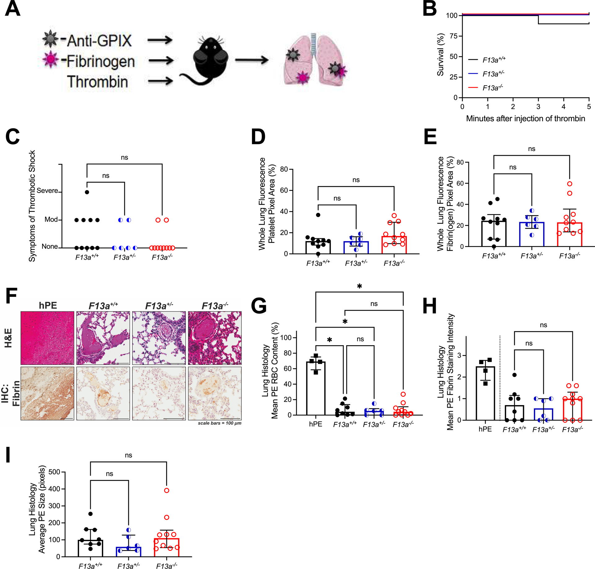
Figure 1. In the thrombin infusion model, F13a+/+, F13a+/−, and F13a−/− mice have similar PE incidence.

(A) F13a+/+ (N=10), F13a+/− (N=6), and F13a−/− (N=10) were injected retro-orbitally with Alexa Fluor-647 labeled fibrinogen and Alexa Fluor-800 labeled anti-GPIX antibodies and then injected intravenously with thrombin and observed. (B) Survival and (C) symptoms of shock. Mice were perfused after the observation period and the lungs were harvested. Fluorescent signal from (D) Alexa Fluor-800-anti-GPIX antibody and (E) Alexa Fluor-647-fibrin(ogen) in perfused lungs. Sections of human PE (hPE) and lung from thrombin-infused mice were analyzed by (F) H&E staining and immunohistochemistry for fibrin (brown staining); scale bars indicate 100 μm. (G) RBCs and (H) fibrin quantified from histology; fibrin staining in hPE is presented for illustrative purposes but not directly compared to mouse PE. (I) Average size of PE in thrombin-infused mice. Statistical comparisons were made by Kruskal-Wallis testing with Dunn’s post-hoc test. Dots represent individual mice; columns and error bars indicate median and interquartile range. *P<0.05; ns, non-significant
