
Figure 2.
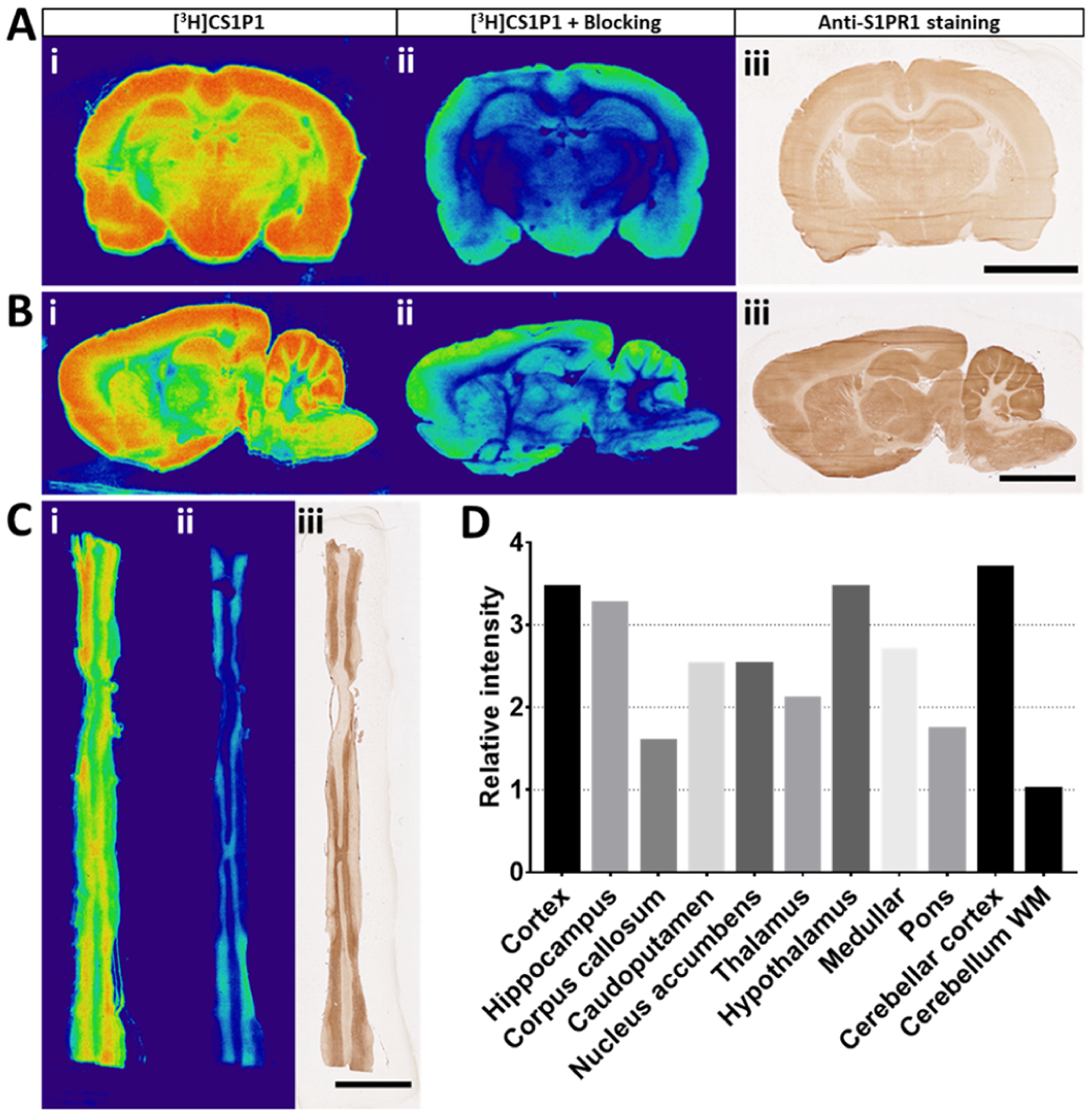
In vitro characterization of [3H]CS1P1 and S1PR1 in CNS of rat. (A–C) In vitro autoradiography of [3H]CS1P1 and immunostaining of S1PR1 in the rat brain and spinal cord. [3H]CS1P1 autoradiograph (i), autoradiograph with NIBR-0213 blocking (ii), and immunostaining (iii) were performed in the rat brain (A,B) and spinal cord (C). Autoradiograph study showed that [3H]CS1P1 is mainly distributed in gray matter with no to very low amount in the white matter in both brain and spinal cord; [3H]CS1P1 can be blocked by S1PR1-specific antagonist NIBR-0213; the distribution of [3H]CS1P1 matched well with immunostaining with anti-S1PR1 (scale bar = 5 mm); (D) relative radioactivity of [3H]CS1P1 in different regions of the rat brain: WM has a relatively low level of bound [3H]CS1P1, whereas GM has a much higher level of bound [3H]CS1P1, among all tested regions, cerebellum WM has the lowest level of bound [3H]CS1P1, and cortex, hypothalamus, and cerebellar cortex show the highest level of bound [3H]CS1P1. GM: gray matter, WM: white matter.