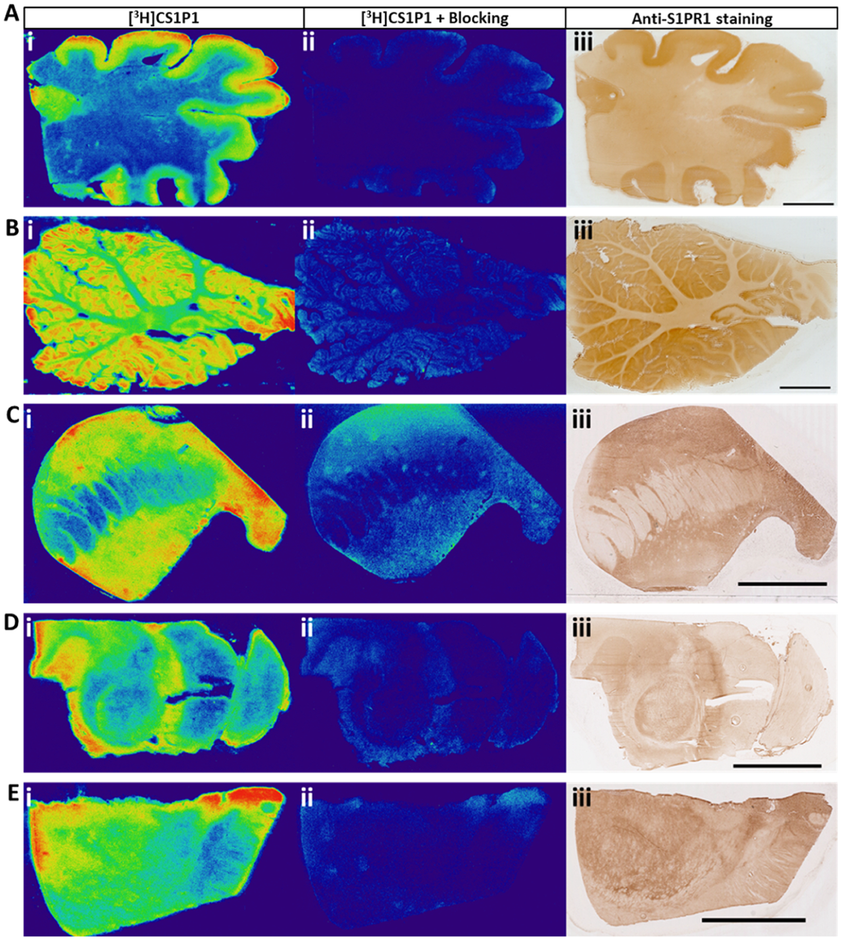
Figure 3.

In vitro autoradiography of [3H]CS1P1 and immunostaining of S1PR1 in the human brain. (A–E) [3H]CS1P1 autoradiograph (i), autoradiograph with NIBR-0213 blocking (ii), and immunostaining (iii) were performed in the human frontal cortex (A), cerebellum (B), striatum (C), midbrain (D), and thalamus (E). Autoradiograph study showed that [3H]CS1P1 is mainly distributed in gray matter with no to very low amount in the white matter; [3H]CS1P1 can be blocked by S1PR1-specific antagonist NIBR-0213; the distribution of [3H]CS1P1 matched well with immunostaining with anti-S1PR1 (scale bar = 1 cm for A and B and 5 mm for C–E).
