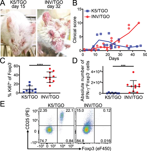
Figure 1: INV/TGO mice developed a lethal autoimmune disease in contrast to the self-resolving disease in the K5/TGO model.
OVA expression in the skin was induced by 1200 μg dox/mL and 0.8–1×10⁶ LN cells from DO11.10 Rag2−/− animals were transferred to K5/TGO or INV/TGO mice. (A) Representative pictures of K5/TGO and INV/TGO mice at the height of disease (15 and 32 days after transfer, respectively). (B) Clinical disease course over the duration of the experiment (representative of 3 experiments, N ≥ 8/group in total). (C, D) sdLNs were analyzed by flow cytometry 22–57 days after transfer. Graphical summary of the Ki67 expression and IFN-γ production by live gated CD4+Foxp3-DO11.10-TCR+ Teff cells. Cumulative data from 4 experiments, N ≥ 8/group. (E) Representative flow plots of Foxp3 and CD25 expression by live gated CD4+DO11.10-TCR+ T cells in the sdLNs of recipient animals 22 days after transfer. Mean and SD is shown.

