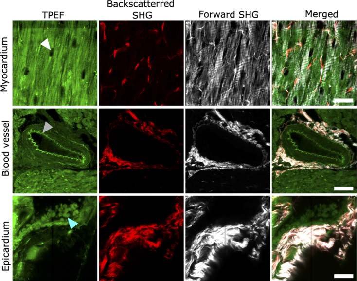
Fig. 1.
Label-free NLOM images of old marmoset cardiac tissue. Representative TPEF, backscattered SHG, forward propagating SHG and merged images are shown for coronary blood vessel, myocardium and epicardial wall from the same heart. TPEF arises from endogenous fluorophores such as blood cells (marked by cyan arrowhead), elastin (marked by grey arrowhead) and other intracellular fluorescent proteins, while the nuclei appear dark (marked by white arrowhead). Backscattered SHG emission is detected mainly from collagen while the forward propagating SHG emission originates with high intensity from both collagen and myosin at the epicardium and myocardium and around the blood vessel. Merged images show signal emission from all channels. Scale bar: 25 µm in all images.

