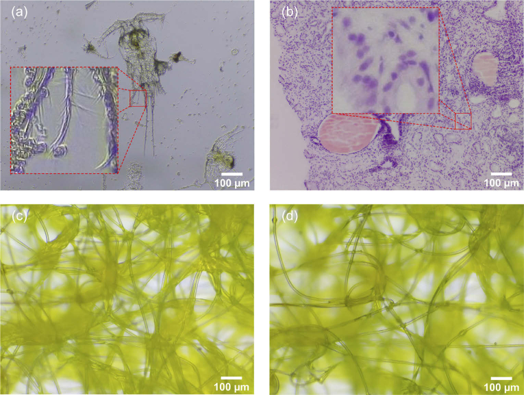
Fig. 7.
RGB raw images captured by μH3D: (a) copepod with bight field transmission illumination and a square section of side 75 μm zoomed in; (b) stained tissue biopsy with trans- and reflective illumination and a square section of side 75 μm; (c) synthetic fibre with transmission bright field illumination; and (d) same fibre with trans- and reflective illumination. These images where captured at size pixels with an achromatic objective 20x, NA 0.4, ∞/0.17.

