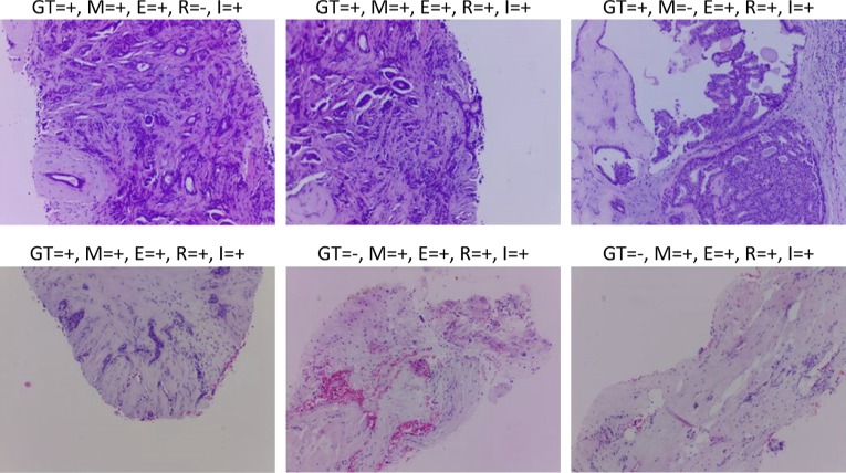
Fig. 10.
Breast tissue samples with Hematoxylin-Eosin staining acquired with μH3D and classified by the four inference models in the server upon client request. Over each sample a legend displays the correspondent values of ground truth (GT) and classification results (i.e., +/- for positive/negative to cancer) for each model: (M)obilenet V2, (E)fficienteNet0 Lite, (R)esNet50, and (I)nceptionV3.

