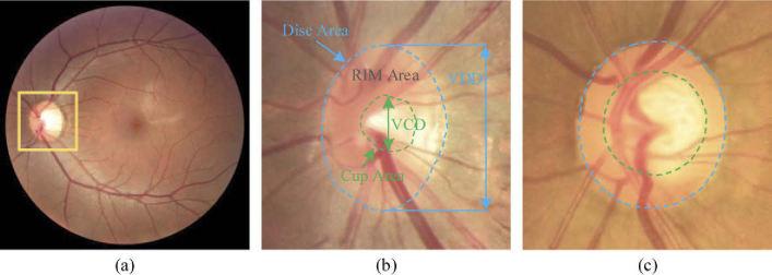
Fig. 1.
Examples of the (a) whole fundus image and the enlarged optic disc area for the (b) normal and (c) glaucoma cases. The optic disc and cup are indicated by the outer blue and inner green dash lines, respectively. VCD and VDD indicate the vertical diameter of the optic cup and disc, respectively.

