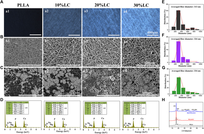
FIGURE 3.
The polarizing optical microscopy (POM) picture (A) and scanning electron microscope (SEM) topography (B) of the PLLA composite scaffold with different LC contents; (C) the response to SEM morphologies and (D) energy dispersive spectrometer (EDS) spectra of poly (L-lactide) (PLLA) and PLLA/LC composite fibers after mineralized in 1.5× simulated body fluid (SBF) solution for 7 days. The fiber diameter distribution of (E) PLLA, (F) PLLA/LC, and (G) hydroxyapatite (HAP)-PLLA/LC. (H) X-ray diffraction patterns of three group samples.

