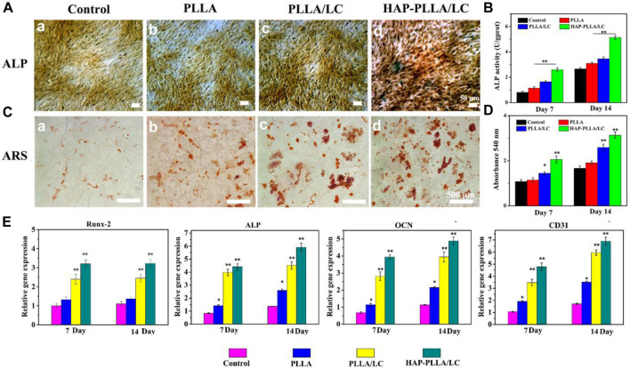
FIGURE 6.
(A) Alkalinephosphatase (ALP) staining and (B) its quantitative analysis of BMSC cells cultured on the scaffolds for 14 days. (C) Alizarin red (AR) staining and (D) its quantitative analysis of BMSC cells cultured on a culture plate, PLLA, PLLA/LC, and HAP-PLLA/LC fiber scaffold for 14 days, respectively. (E) Real-time quantitative PCR (RT-qPCR) analysis of osteogenic and vascularized-related gene expression of cells after culturing for 7 and 14 days.

