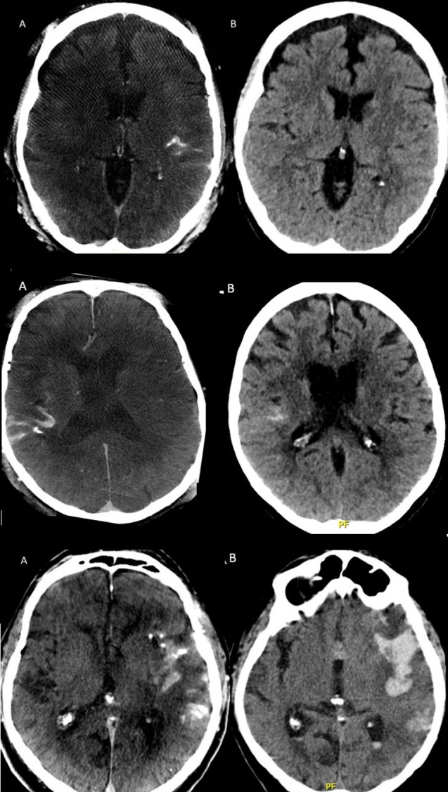
Figure 2.

Column A (left) and B (right) displaying CBCT immediately after thrombectomy and regular CT scan after 24 hours, respectively. Upper row: minimal contrast agent extravasation in the left sylvian fissure initially with complete resolution on follow-up. Middle row: moderate contrast agent extravasation in the right sylvian fissure with marked but incomplete resorption at 24 hours (rated as asymptomatic intracranial bleeding). Lower row: important contrast agent extravasation in the left sylvian fissure with an associated temporal parenchymal hemorrhage and intraventricular extension on the control CT the following day (rated as symptomatic intraparenchymal hemorrhage). CBCT=flat panel cone-beam CT.
