Figure 5.
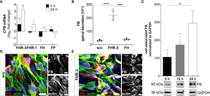
FHR-3 increased FB expression and secretion in ARPE-19 cells. (A) CFB mRNA increased after apical FHR-3 treatment of ARPE-19 cells, but not following incubation with FHR-1, FH or FP. This effect could be confirmed at the protein level: (B) Apical FB secretion was increased 24 h after FHR-3 incubation. (C) Western blots of ARPE-19 cell lysates detected a time-dependent increase in FB levels (95 kDa) 24 h after FHR-3 treatment. Supplementary Figure 1L shows full Western blots. (D, E) Increased FB protein levels were detected by immunofluorescence using specific anti-FB (red) and anti-actin (green) antibodies 12 h after FHR-3 treatment. FB was co-localized partly with actin stress fibers (yellow). Scale bars 40 µm. (A–C) w/o untreated control (dotted line). (A–C) Mean with standard deviation is shown. ****p < 0.0001, **p < 0.01, *p < 0.05. (A) Wilcoxon matched-pairs signed rank test (n = 3); (B, C) Ordinary one-way ANOVA, Turkey’s multiple comparisons test (n = 3).

