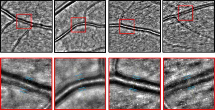
Figure 2.
AO fundus camera exemplary images. The upper row shows 4° × 4° images of a first-order arteriole with a marked region of interest within a red square. The lower row shows the magnification of the red square from the reference image above with blue lines highlighting the vessel wall borders. They show asymmetric vessel wall diameters in this cardiovascular risk population.

