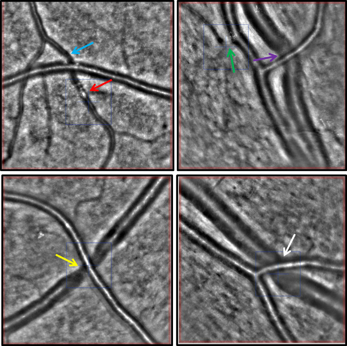
Figure 3.

AO fundus camera exemplary images (4° × 4° image sections). The blue and green arrows mark sites of focal arterial narrowing. The red arrow highlights an intraluminal hyperreflective material. The purple, yellow, and white arrows mark sites of AVN at arteriovenous crossing sites.
