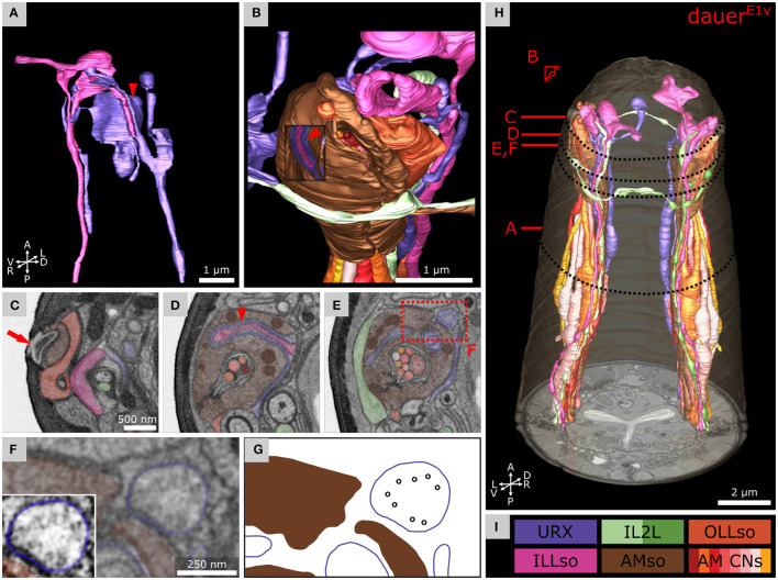
Figure 5.
Anterior dendritic endings of dauer URX neurons are putatively ciliated and extend distal structures to enclose ILLso and to be enclosed by AMso support cells. (A) Volumetric 3D reconstruction of URXL sensory ending enclosing a branch of ILLsoL (arrowhead). As an example, the left side is shown. (B) Example of the spatial arrangement of URXL, ILLsoL, AMsoL, OLLsoL, and IL2LL. Part of AMsoL is clipped to allow a view inside. AMsoL encloses the part of URXL which encloses the branch of ILLsoL shown in A (arrowhead). OLLsoL encloses AMsoL at the amphid opening. The crown-like dendrite of IL2LL is in direct contact with AMsoL. (C) Transverse FIB-SEM section at the amphid opening (arrow) in the cuticle, which marks the beginning of the amphid channel formed by AMsoL. AMsoL is enclosed by OLLsoL. (D) Transverse section at the position where AMsoL encloses URXL which encloses ILLsoL (arrowhead). (E) Transverse section at the position where AMsoL encloses URXL but without enclosing ILLsoL. The IL2LL branch passes AMsoL at the periphery. (F) Higher magnified part of E showing microtubules inside URXL. Inset shows the same image with enhanced contrast. (G) Scheme indicating microtubules in F. (H) Overview of reconstruction of all mentioned cell endings. (I) Names and color code of cells shown.

