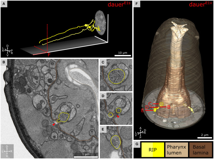
Figure 7.
Anterior dendritic endings of RIP neurons of dauer enter the pharyngeal nervous system. (A) Tracing of RIP neurons. (B) Transverse FIB-SEM section at the position where the ending of RIPR is entering the pharyngeal nervous system through an opening of the pharyngeal basal lamina. (C) Bouton with electron dense vesicles of RIPR inside the pharyngeal nervous system. (D) RIPR split at the pharyngeal basal lamina just before its ending is entering the pharyngeal nervous system. (E) Transverse section posterior of B where RIPR forms a small bouton filled with electron dense vesicles. (F) 3D reconstruction of RIP endings projecting through the pharyngeal basal lamina. (G) Names and color code of cells and structures shown.

