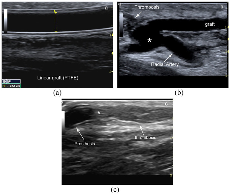
Figure 3.
B-Mode image of an arteriovenous graft. Typical grey scale image of a linear radial-cephalic antecubital linear graft (double-track sign) (a). Loop graft are usually placed within the forearm and anastomosed to the brachial artery and the basilic or brachial vein. Linear bridge in PTFE between antecubital radial artery and basilic vein after the thrombosis of vein draining proximal radio-cephalic fistula (*anastomotic chamber) (b). Intraluminal thrombotic material downstream of the venous anastomosis between prosthesis (*) and native cephalic vein (c).

