Fig. 5.
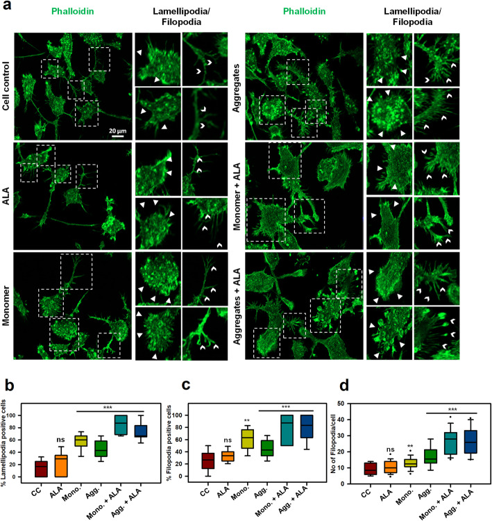
Enhancement of actin-rich structures for migration in microglia. Actin plays a significant role in the migration of microglia by providing mechanical strength as well as direction support. a Actin based structures lamellipodia, filopodia were observed after 24 h exposure Tau monomer, aggregates along with ALA to microglia cells by fluorescence microscopy. The enlarge panel indicates the presence of lamellipodia (white triangles), filopodia (white arrow heads) from the fluorescence image. The enlarged regions are denoted with the white dotted square in the image, scale bar is 20 μm. b The graph represents quantification of percentage of lamellipodia positive cells per 10 fields of different treatment groups, (p < 0.001) as compared to cell control (untreated) and ALA. c The graphical representation of percentage of filopodia positive cells per 10 fields of different treatment groups Tau monomer, aggregates along with ALA showed high significance of (p < 0.001) d Quantification of number of filopodia extensions present per cell per 10 fields of different groups, significance is (p < 0.001). The significance was analyzed with Tukey’s Kramer, significant when mean difference between treatment groups (X-X’) > T (Tukey’s criteria). The annotations used in graph are as follows- CC (Cell control), ALA (α-linolenic acid), Mono. (monomer), Agg. (aggregates), Mono. + ALA (monomer plus ALA), Agg. + ALA (aggregates plus ALA)

