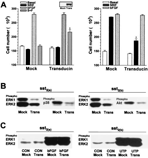
FIG. 8.
Involvement of βγ subunits in the mediation of the proliferative and phosphorylation effects induced by somatostatin in CHOsst2(a) and CHOsst2(b) cells. (A) The effect of transient expression of the βγ sequestrant transducin on cell proliferation induced by 100 nM somatostatin (SRIF; solid histograms), 10 ng of bFGF per ml (hatched histograms), or somatostatin in the presence of bFGF (S+F; shaded histograms), determined 24 h following application to partially denuded monolayers of either CHOsst2(a) (left graph) or CHOsst2(b) (right graph) cells is shown. The effect of transfection with the empty plasmid, pCDNA3, is represented by the histograms labeled Mock, and open histograms show basal repopulation. Values are expressed as the mean cell number harvested from a single coverslip (from two separate transfections, three replicates). Groups labeled with an asterisk are significantly different from the respective treatment group for cells without transducin expression (P < 0.05). Cell samples extracted immediately prior to partial denudation that had been transfected 48 h previously with either pCDNA3 (Mock) or pCDNA3 incorporating transducin cDNA (Trans) were analyzed by Western detection using an appropriate antibody to confirm expression of transducin (inset). (B) The effect of βγ sequestration on somatostatin-induced phosphorylation of ERK1, ERK2, and p38 in CHOsst2(a) cells or of ERK1, ERK2, and Akt in CHOsst2(b) cells. Western detection was performed using phosphospecific antibodies of samples prepared from whole-cell extracts of partially denuded monolayers incubated in the presence of 100 nM somatostatin for 10 min. Samples from cells transfected with the empty plasmid 48 h prior to partial denudation are labeled Mock, and those from cells expressing transducin are labeled Trans. (C) The effect of βγ sequestration on the phosphorylation of ERK1 and ERK2 stimulated by 10 ng of bFGF per ml in CHOsst2(a) cells or by 100 nM UTP in CHOsst2(b) cells following incubation for 10 min immediately after partial denudation of confluent monolayers. Western detection was performed using phosphospecific anti-ERK antibodies of samples prepared from whole cells that had been either transfected with the empty plasmid 48 h prior to partial denudation (Mock) or transfected to overexpress transducin (Trans).

