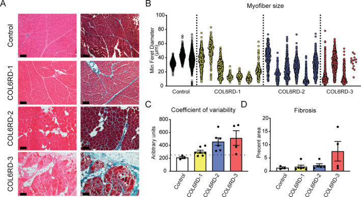
Figure 1.

Muscle histologic group classification and morphometry. (A) Representative hematoxylin & eosin and Gömöri trichrome staining of frozen muscle sections used to qualitatively stratify the biopsies into three histologic severity groups: COL6‐RD‐1, COL6‐RD‐2, and COL6‐RD‐3. (B–D) Morphometric quantification of fiber size (B), fiber size variability (C), and fibrosis (D) of a subset of biopsies confirm the expected trends of histologic severity and qualitative classification. Note the severe myofiber atrophy in COL6‐RD muscle biopsies (B). The dotted line in panel C indicates the cutoff for normal coefficient of variability (250). The error bars represent standard error of the mean. Scale bar = 50 μm.
