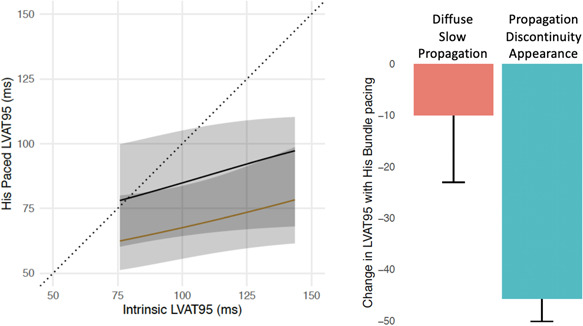
Figure 3.

Impact of propagation discontinuity during intrinsic conduction in LBBB on change of LVAT with HBP. (Left) Ordinal regression of HBP LVAT‐95 from intrinsic LVAT‐95 in patients with LBBB. The black line represents patients with diffuse slow propagation (absence of propagation discontinuity) in LBBB. The yellow line represents patients with regions of propagation discontinuity in LBBB. Patients with propagation discontinuity were more likely to achieve a shorter LVAT‐95. (Right) change in LVAT‐95 from intrinsic LBBB to HBP in patients without (red) and with (blue) appearance of propagation discontinuity in LBBB. HBP, His bundle pacing; LBBB, left bundle branch block; LVAT‐95, left ventricular activation time of 95% of activations
