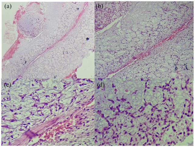
Figure 2.
Histological findings of the first biopsy: a lobular tumor, with peripheral hypercellularity. Cells are mildly pleomorphic, spindled to stellate-shaped. Nuclei are elongated, with diffuse chromatin, inconspicuous nucleoli, and low mitotic activity. (a): HE × 40B: HE × 100, (b) HE × 400, and (c) HE × 400.

