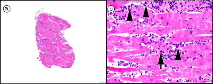
Figure 1.
Endomyocardial biopsy showing a polymorphous (mixed) active myocarditis with lymphohistiocytic inflammation and increased eosinophils (b, arrowheads). Myocyte injury was diffuse throughout the sampled tissue (b, arrow). No giant cells were identified throughout extensive sectioning of the biopsy, but vaguely granulomatous inflammation was apparent. (a, 40× original magnification, and b, 400× original magnification; both hematoxylin and eosin stain).

