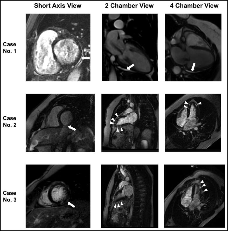
Figure 2.
Cardiac magnetic resonance imaging of Cases 1–3 in short axis (first column), two-chamber (second column), and four-chamber (third column) views. Case 1: Mild late gadolinium enhancement (LGE) is seen in the inferolateral region in the pericardium in all views (arrow). Case 2: LGE is seen in the mid wall region in a multifocal distribution in all views (arrows and arrowheads). Case 3: LGE is seen in the inferolateral and lateral wall in all views (arrows and arrowheads).

