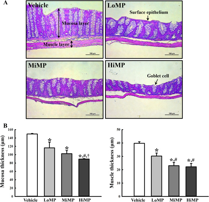
Fig. 1.
Histopathological structures of the mid colon. A H&E stained sections of the mid colon from the Vehicle, LoMP, MiMP, and HiMP treated groups were observed at 200 × magnification using an optical microscope. B The histopathological parameters were determined using the Leica Application Suite. Four to six mice per group were used to prepare the H&E stained slides, and the histopathological parameters were measured in duplicate for each slide. The data are reported as the mean ± SD. *p < 0.05 compared with Vehicle treated group; #p < 0.05 compared with LoMP treated group; †p < 0.05 compared with MiMP treated group. Abbreviation: LoMP, Low concentration of microplastics; MiMP, Medium concentration of microplastics; HiMP, High concentration of microplastics

