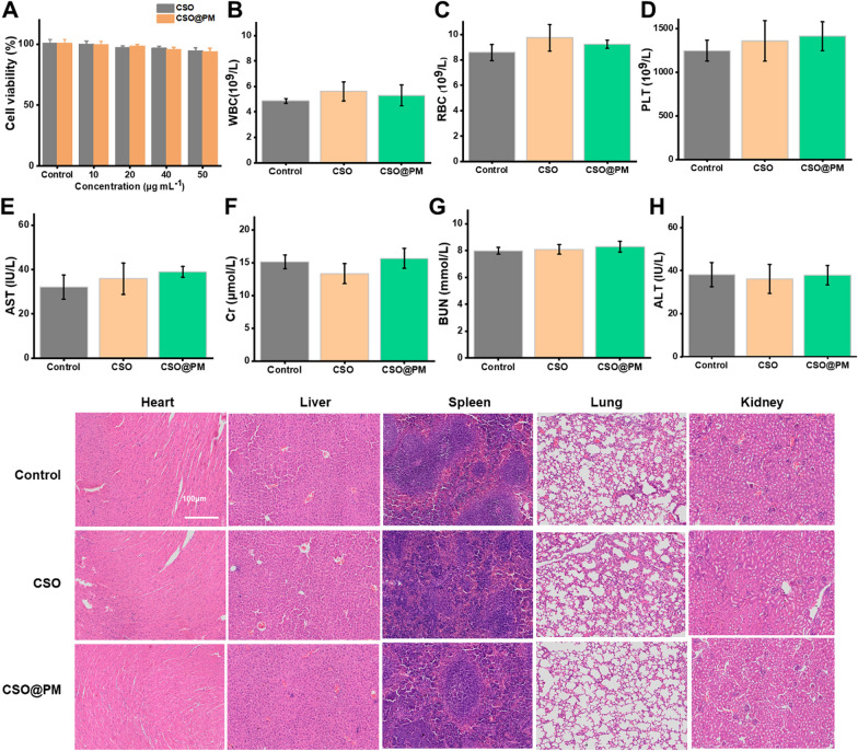
Fig. 7.
In vitro and in vivo biocompatibility of CSO@PM. A Cell viabilities of 3T3 fibroblasts treated with different concentrations of CSO@PM. B–H Hematology and blood biochemistry analysis results for healthy Balb/c mice sacrificed 21 days after intravenous injection with CSO@PM at a concentration of 500 mL−1 (n = 3). PBS-treated mice were used as controls. B White blood cells, C red blood cells, D platelets, E aspartate aminotransferase (AST), F creatinine (Cr), G blood urea nitrogen (BUN), and H alanine aminotransferase (ALT). I H&E staining images of the major organs (heart, liver, spleen, lung, and kidney) from mice 21 days after intravenous injection with CSO@PM (500 μg mL−1). Scale bar: 100 μm

