Fig. 3.
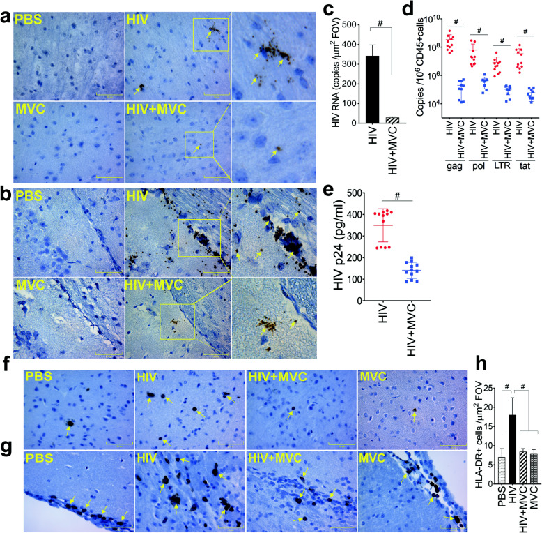
MVC decreased viremia and abrogated HIV-induced cellular infiltration in the brain of infected animals. Brain HIV RNA copy (yellow arrows) numbers were quantified by RNAscope and, for each experimental group, representative images from the somatosensory cortex (a) and meningeal / somatosensory area layer 1 (b) are shown. c: Metamorph software was used to quantify HIV RNA copies numbers in all samples. For each animal brain sample, 10 random fields-of-view (FOV) were analyzed (5 FOV from the somatosensory cortex and 5 FOV from the meningeal / somatosensory area layer 1). d: Levels of HIV-1 gag, pol, LTR, and tat genes in brain tissues were quantified by qPCR and normalized to samples’ hCD45+ cells levels. e: HIV-1 p24 antigen levels in plasma samples were quantified by ELISA. f, g: HLA-DR expression in brain tissues was analyzed by immunohistochemistry and, for each experimental group, representative images from the somatosensory cortex (f) and meningeal / somatosensory area layer 1 (g) are shown. h: Metamorph was used to quantify HLA-DR+ cells in all brain samples and for each sample, 10 random FOV (5 FOV from the somatosensory cortex and 5 FOV from the meningeal / somatosensory area layer 1) were analyzed. For panels a, b, f, and g, images were at 40X. The four animal groups included PBS, HIV, HIV + MVC, and MVC; 9 to 12 animals in each group. #P < 0.0001. Error bars represent SD

